Note: MRI images are flipped, meaning what you see on the left is actually on the right side of the brain and vice versa. We selected these images as carefully as we could, but they aren’t shots of exactly the same location in exactly the same position. Moreover, the images were taken on different equipment using different contrast fluids, which is why the tumor seems brighter on one image than on another. Last but not least, reading MRI images is an art and a science; we hope these images are helpful to you on your journey, but please don’t use them to draw conclusions about your own MRIs. You can click on each image for a larger view.
Back view
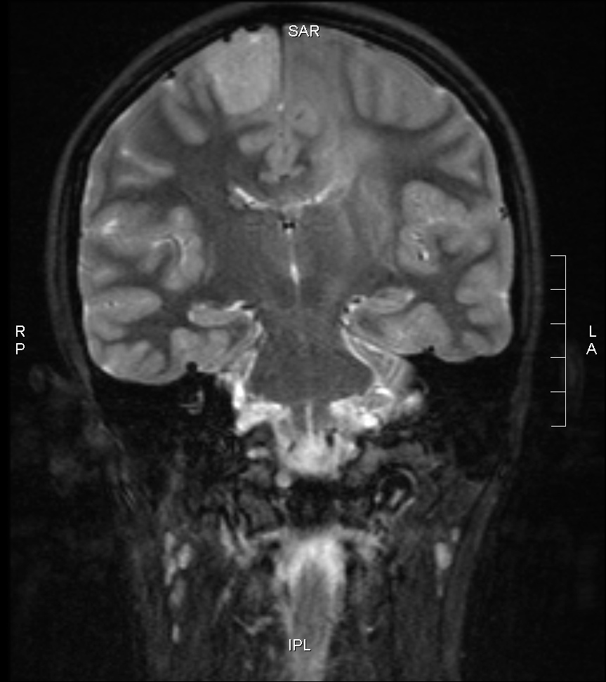
Sadie’s first MRI. You can see the large solid mass at the top right (top left in the image) and the smeary area in the left hemisphere (right in the image). This smear is what made Sophie’s tumor inoperable; it’s like a ribbon around her neural pathways. You can’t see it in this image, but the top right mass and the left smear are connected.
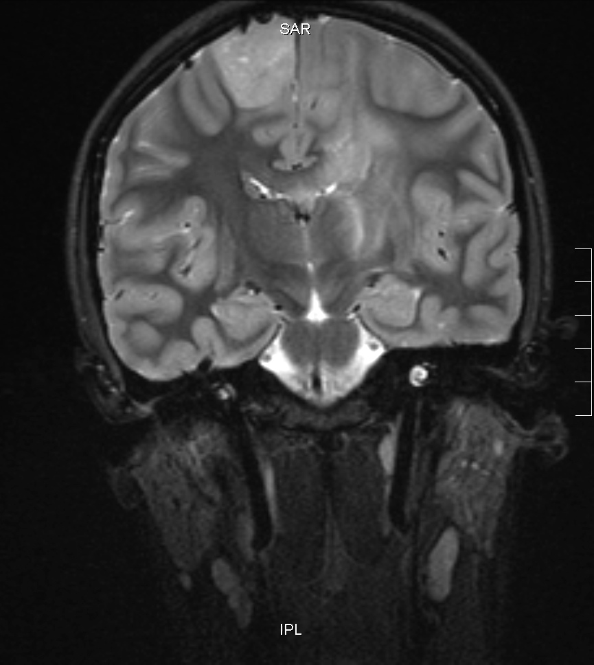
Three weeks later, you can see that the top right mass has grown slightly and the left smeary area has grown significantly.
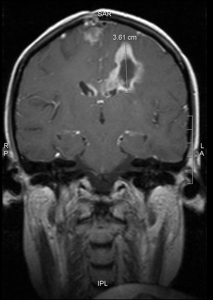
Sadie’s first post-treatment MRI. Radiation and chemotherapy have kicked butt on the top right tumor; it’s shrunk dramatically. Unfortunately, the smeary areas are still quite large. The dark center in the left tumor area indicates necrosis (dead tissue), which could be from radiation; it’s also a hallmark of glioblastoma multiforme (GBM): a GBM grows so quickly its blood supply can’t keep up with it, and the center dies off.
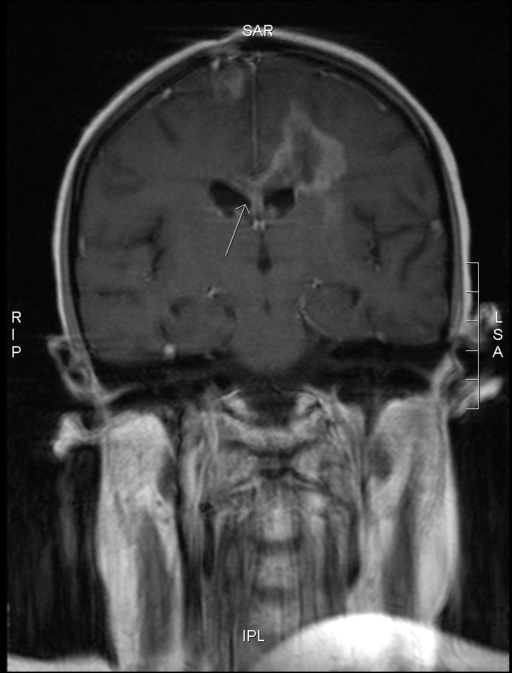
Six weeks later, another MRI shows the top right mass to be stable. We are devastated to hear that the tumor in the left hemisphere has grown. (It’s hard to see from these single images, but that’s what the radiologist told us.) Note also that the tumor has crossed the midline (see the arrow). Sadie starts cisplatinum-etoposide treatment 11 days later, as soon as her blood is good enough.
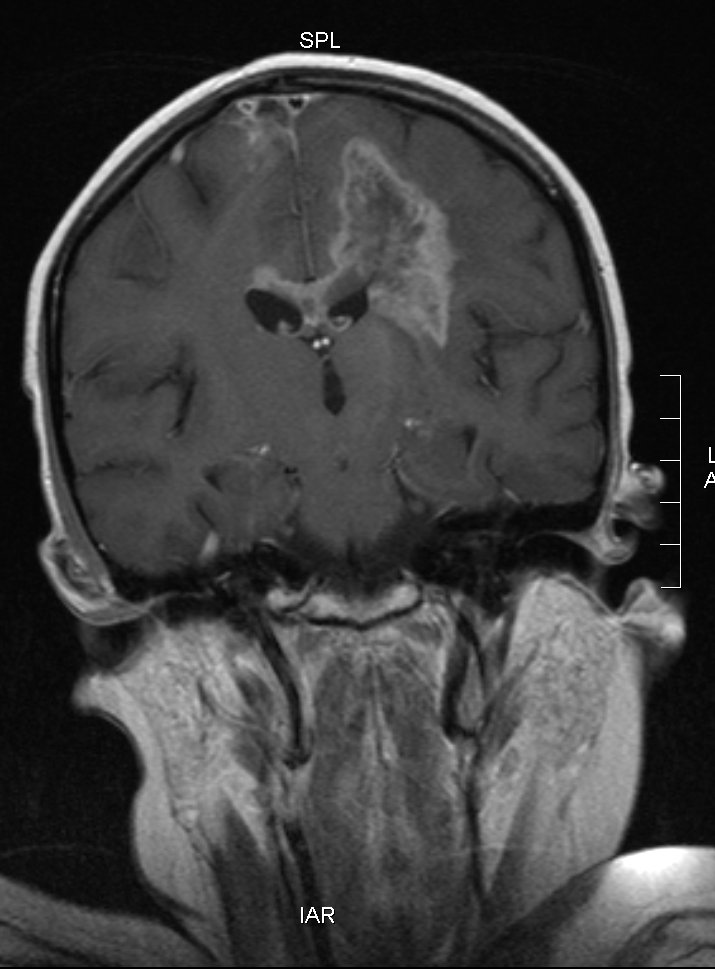
Another six weeks later, the tumor has grown dramatically. We decide to stop treatment.
Side view
These series of images show changes to the tumor in the left hemisphere between August 10 (top series) and September 18 (bottom series), 2006. Each series starts at the midline in the brain (the dividing line between left and right hemispheres) and proceeds out toward Sadie’s left ear. Note that the August 10 scans begin slightly closer to the midline (you can see Sadie’s spinal cord in the first image) than the September 18 scans. This means each August 10 image comes “before” (closer to the midline than) its corresponding September 18 image.
You can right-click on each image and open a larger version in a new window if you want to compare the two series.


The new brain stem tumor
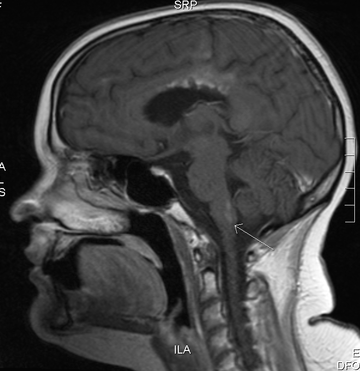
Contrary to all expectation, the existing two tumor sites have not changed since the last MRI on September 18. But there’s a new kid in town: the arrow in the image points to the new lesion on Sadie’s medulla oblongata. The medulla oblongata is responsible for heart rate, breathing, swallowing, and vomiting and controls several skeletal muscles involved with speech.